
 +86 18928918121
Siemens MAGNETOM Vida RT Pro 3.0T with Motion-Perception Simulation & Positioning
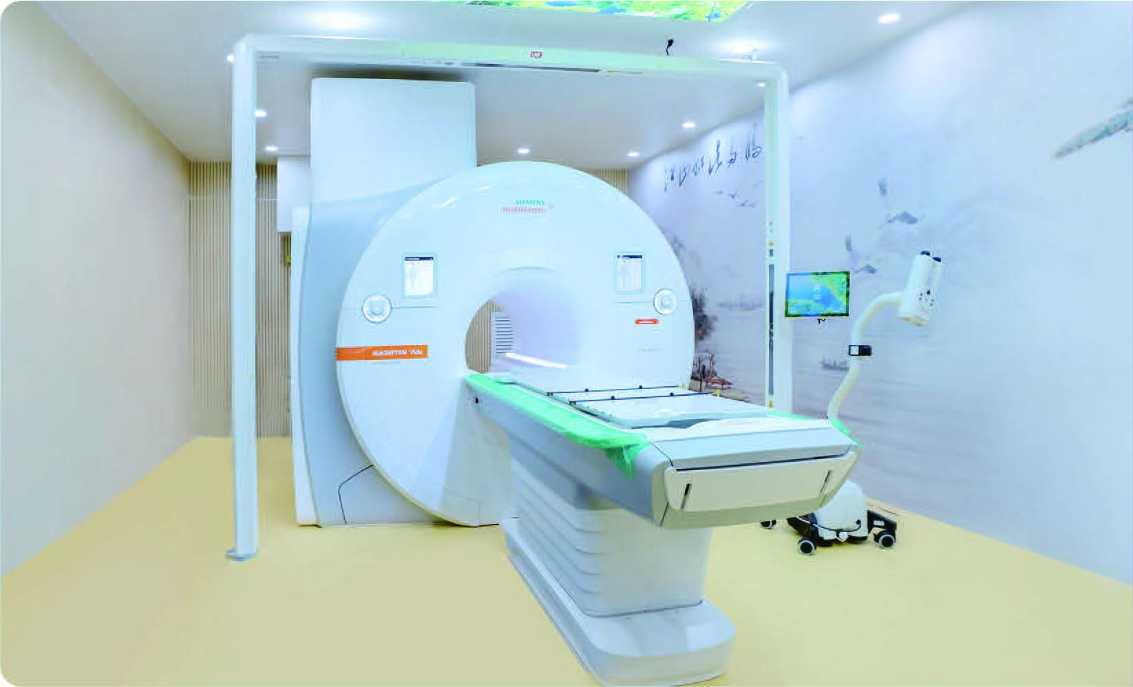
MRl Simulation Makes Radiation Therapy More Precise.
√Silent Scan
√Accurate Localization
√Intelligence and Effciency
√Enhanced Comfort, Reduced Noise, and Accelerated Scanning
√Smarter, More Efhcient, and Easier to Use
Siemens Vida MRI-based Synthetic CT Technology
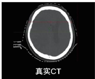
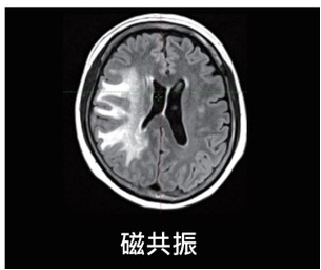
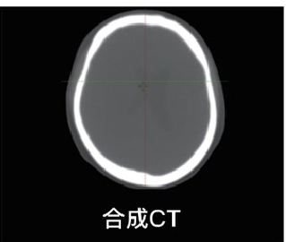
Siemens’MR-only radiotherapy solution enables direct generation of “synthetic CT” images from high-resolution MRI data, completely eliminating radiation exposure. This approach is particularly valuable for pediatric, pregnant patients, and those requiring repeated treatment courses.
With a single MRI scan, clinicians achieve both precise target delineation and accurate radiation dose calculation, removing the need for CT simulation and streamlining the entire radiotherapy workflow. This technology is primarily implemented for tumors in the head and pelvic regions.
Our Hospital’s First MRI-based Synthetic CT Case -May 28, 2025
Self-gated 4D MRI for respiratory motion management
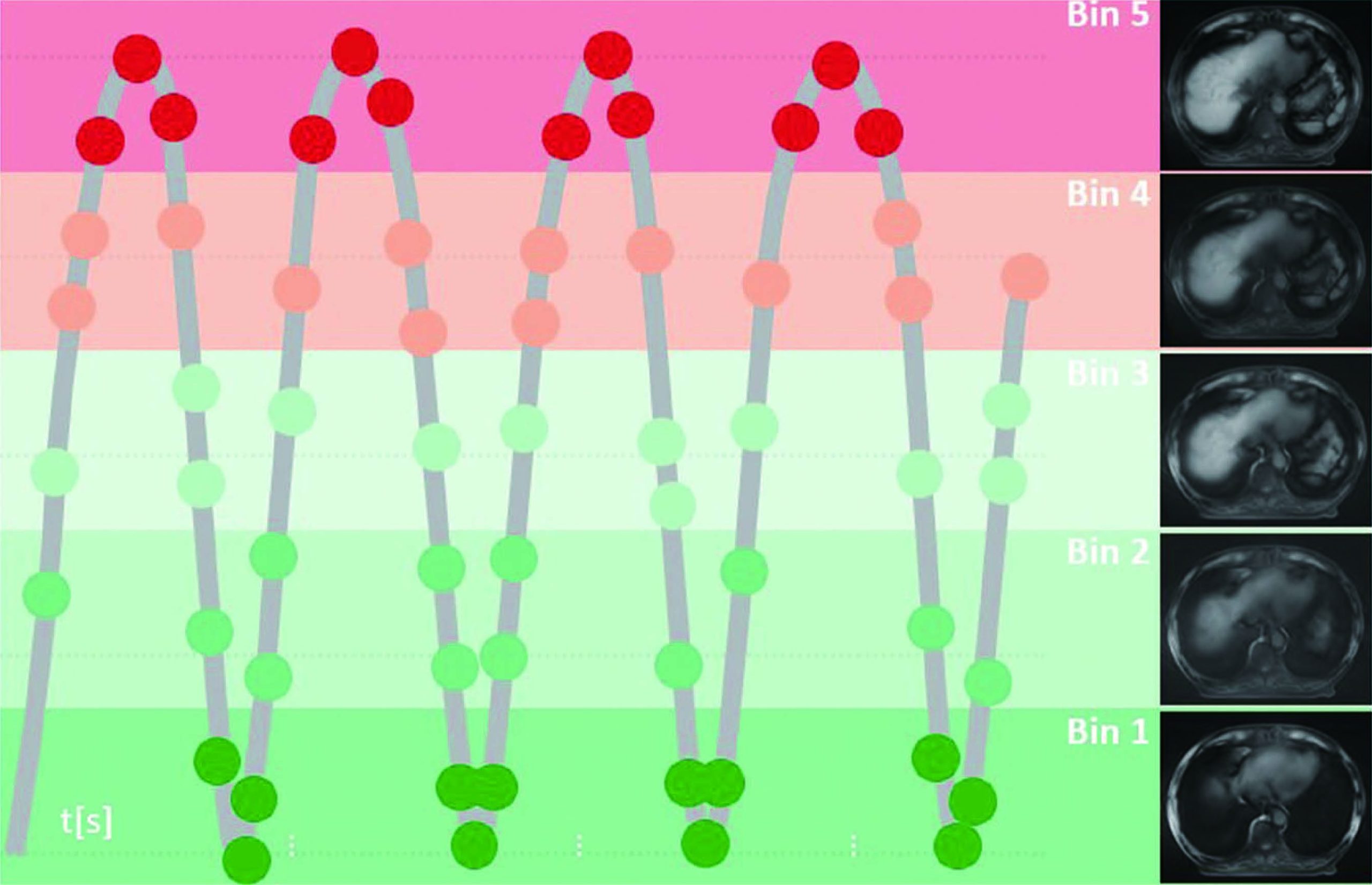
Motion artifacts in 3D imaging often occur due to physiological movements such as respiration, cardiac activity, peristalsis, and muscle contractions. While rapid imaging techniques like single-slice CT may capture brief moments of the respiratory cycle, inconsistent phase sampling across slices frequently introduces motion distortions.
Siemens Healthineers addresses this with 4D MR-RT Self-Gating Technology, which dynamically tracks abdominal and thoracic organ motion throughout acquisition. This innovation eliminates external sensors while maintaining exceptional soft-tissue contrast and phase-consistent free-breathing sorting.
The technology enables full free-breathing radiotherapy simulation, eliminating breath-holds to enhance patient comfort. By suppressing motion artifacts and improving spatial accuracy, it achieves superior tumor targeting precision and facilitates accurate delineation of moving targets and organs at risk.
Our Hospital’s First Clinical Application of 4D MRI Gating Technology – July 4, 2025
Distributed Nuclear Magnetic Adaptive Technology
Prior to treatment, an MRI simulation scan is performed to re-con和m the tumor’s position and morphological changes. The radiation oncologist then modifies the target volume accordingly. A rapid planning process is used to re-optimize the treatment plan. Finally, the patient is transferred to the linear accelerator via a shuttle couch, which ensures their position remains unchanged throughout the process.
Key Features of Distributed Nuclear Magnetic Adaptive Technology
More Precise Image Guidance: Compared to current CBCT -guided adaptive radiotherapy, MRI provides images free from air-bone artifacts, offers superior soft-tissue resolution, and delivers no additional radiation dose.
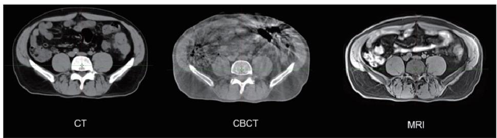
Faster Treatment Delivery: Compared to existing integrated MR曰inear accelerators (MR-linacs), which require approximately one hour for a single online adaptive session, the distributed NMR adaptive radiotherapy system significantly reduces the treatment time. The entire process, including a 6-minute MRI scan and a 6-minute plan optimization, is streamlined to be completed within 30 minutes.
Enhanced Operational Efficiency: Unlike existing adaptive radiotherapy systems that must pause treatment after imaging to wait for target modification and plan re-optimization, the distributed adaptive approach efficiently coordinates the MRI simulator with the treatment accelerator, eliminating machine idle time.
Our Hospital’s First Clinical Application of Distributed Nuclear Magnetic Adaptive Technology – September 22, 2025
Adaptive Radiotherapy Workflow Based on MR Simulator for Tumor Segmentation

The patient lies flat on the air mattress in the correct position.

Initiate the MR device to acquire images.

After the scan is completed, the air compressor is activated, causing the air mattress MR platform.

The patient is gently and smoothly transferred from the MR system platform to the transport cart

During the patient transfer from the simulation CT to the linac, the physicist imported the acquired MRI images into the treatment planning system.

The patient is transported from the MR simulation suite to the radiation therapy vault via a transport cart by the therapist.
Using daily imaging to assess displacements of the target and OARs, the radiation oncologist redefined the target volume in approximately five minutes.
During transport, the patient must remain calm and avoid turning or moving to prevent displacement of the tumor.
The physicist conducts a dose re-optimization for the revised target volume and completes the handover by sending the new plan to the treatment linac and notifying the radiation therapist.
The patient is smoothly transported to the radiotherapy room via the cart and then carefully slid horizontally onto the treatment couch.

Treatment is initiated with the adapted plan once the new plan has been uploaded and the patient is correctly positioned on the treatment couch.
The complete fractional adaptive radiotherapy process takes under 25 minutes. Patients daily receive a treatment plan adapted to their real-time anatomical changes.
GE SIGNATM Premier 3.0T MRI System
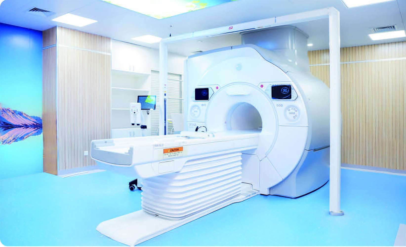
Ultra-high-end medical imaging platform
√Ultra-High-Resolution Diffusion lmaging
√Ultra-High-Accuracy Functionallmaging
√Enabled Significantly Faster Scan Times
The Advantages of SIGNA Premier for Precision Radiotherapy Simulation and Localization
More professional
The high-performance, form-fitting exclusive blanket-style AIR Coil delivers unmatched image quality.The Premier model incorporates a new coil material with an INCA fiber loop design, marking a revolutionary breakthrough that makes MRI coils lighter and more flexible. The lightweight, high-channel design of the AIR Coil simplifies radiotherapy patient positioning and sign巾cantly improves the visualization of small lesions and tissue boundary sharpness, achieving optimal imaging quality.
More efficient
It is the globally unique MRI system capable of achieving an 80 mT/m gradient field strength within a large-bore design. Combined with an internationally leading 148-channel RF platform, it enables high-channel-count imaging for faster scan speeds. A complete set of standardized radiotherapy simulation and localization scanning protocols, covering all body regions with standardized sequences and post-processing, delivers superior imaging efficiency.
Skillful
The system features a specialized deformation control design with torque-balanced gradient damping. It combines an industry-best, large-bore magnet with superior field homogeneity and an exclusive 3D gradient deformation correction technology (Gradwrap). This consistent, minimal-deformation steady-state gradient design effectively overcomes the inherent geometric distortions of MRI, delivering more accurate image data and providing highly precise spatial localization for accurate radiotherapy.
发表回复